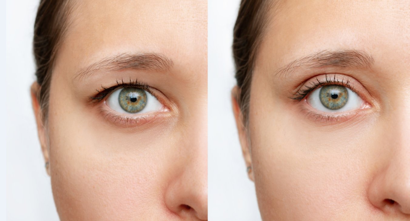
The notion of surgery around the eyes usually conjures images of purely aesthetic improvements, focused on diminishing the visual hallmarks of aging such as under-eye bags or skin laxity. However, to frame eyelid surgery—medically termed blepharoplasty—solely within the realm of cosmetic enhancement fundamentally misunderstands its profound functional capacity. For a significant number of individuals, particularly as they advance in age, the progressive drooping of the upper eyelid tissue, a condition known as dermatochalasis, transitions from a matter of appearance to a tangible, measurable obstruction of sight. This gradual encroachment upon the visual field, an insidious narrowing of the world’s periphery, can severely compromise the ability to perform routine activities, turning simple tasks like driving or reading into efforts of constant, straining compensation. Eyelid surgery in these specific, medically documented instances is not a luxury; it represents a functional imperative aimed directly at restoring unimpeded vision and alleviating the chronic symptoms associated with constant ocular effort.
The progressive drooping of the upper eyelid tissue… transitions from a matter of appearance to a tangible, measurable obstruction of sight.
The mechanics of vision impairment caused by excessive upper eyelid skin are rooted in simple physics. The natural process of aging leads to a loss of elasticity and the stretching of the thin skin and supporting musculature of the eyelids. This redundant tissue eventually creates a heavy fold, which descends and rests directly on the lash line, or in more severe cases, hangs below it. This physical barrier immediately clips the superior and superior-temporal visual field, the very areas that are critical for noticing high-mounted traffic lights, seeing objects in the lane next to you, or maintaining a continuous line of text while reading. This is not a perceived issue; it is a demonstrable deficit that specialists quantify with objective testing. For the patient, the daily consequence is a perpetual sense of heaviness over the eyes, a habitual tilting of the head backward, or the constant, subconscious use of the forehead muscles to lift the eyebrows in a desperate attempt to hoist the obstructing skin out of the sight line.
This redundant tissue eventually creates a heavy fold, which descends and rests directly on the lash line.
Distinguishing between purely aesthetic blepharoplasty and a functional procedure is critical, as it dictates the entire clinical pathway, including the potential for medical coverage. Functional blepharoplasty is specifically performed to address vision-related problems, chief among them the removal of excess skin or fat that physically blocks vision. An even more specialized procedure, known as ptosis repair, addresses the underlying weakness or detachment of the levator muscle, which is the primary muscle responsible for lifting the upper eyelid margin itself. When this muscle is weakened, the eyelid margin droops over the pupil—a true eyelid droop, or ptosis—which represents a more complex functional deficit than mere excess skin. An oculoplastic or ophthalmic surgeon is uniquely qualified to diagnose the precise origin of the obstruction, determining whether the problem is simply redundant skin (dermatochalasis) or a muscular issue (ptosis), or more commonly, a combination of both.
Functional blepharoplasty is specifically performed to address vision-related problems, chief among them the removal of excess skin or fat that physically blocks vision.
The cornerstone of documenting the medical necessity for this type of eyelid surgery lies in specialized diagnostic testing. The most common and essential of these is the automated visual field test, a standardized procedure that maps the extent of the patient’s peripheral vision. This test is performed twice on each eye: once with the upper eyelid left in its natural, drooping state, and then a second time with the excess eyelid skin gently taped up or lifted out of the field of vision. The resulting comparison provides an objective, numerical measurement of the visual field loss directly attributable to the eyelid tissue. Many medical insurance providers, including governmental programs, mandate a specific degree of measured improvement—often a minimum percentage increase in the superior visual field when the lid is lifted—to approve the procedure as medically necessary rather than cosmetic. This stringent criterion ensures the surgery is truly focused on sight restoration.
The most common and essential of these is the automated visual field test, a standardized procedure that maps the extent of the patient’s peripheral vision.
Patients who strain against their drooping eyelids develop a cluster of compensatory symptoms that extend beyond the initial visual field loss. The perpetual engagement of the forehead muscles to maintain an elevated brow line frequently leads to chronic tension headaches, often described as an ache that starts above the eyebrows and radiates across the forehead. Furthermore, the constant effort to see underneath a heavy lid causes significant eye fatigue, especially during visually demanding activities like extended computer use, reading, or driving at night. By successfully removing the visual obstruction through functional blepharoplasty or ptosis repair, the surgeon effectively eliminates the need for this compensatory muscular effort. Patients frequently report that the primary benefit is not just a wider field of view, but a profound relief from the chronic strain and the cessation of these daily headaches, which underscores the interconnected nature of the ocular and muscular systems.
The perpetual engagement of the forehead muscles to maintain an elevated brow line frequently leads to chronic tension headaches.
The surgical procedure itself, particularly for the upper eyelid, is a delicate and precise undertaking, often performed in an outpatient setting under local anesthesia with sedation. The surgeon carefully plans the incision to be concealed within the natural crease of the upper eyelid, a detail that maintains the aesthetic integrity while fulfilling the functional goal. Through this incision, the necessary amount of redundant skin, and sometimes a small amount of underlying fat and muscle, is meticulously excised. If the diagnosis includes ptosis, the surgeon will also proceed to tighten or reattach the levator muscle to restore the correct height and contour of the eyelid margin. The precision of this technique is paramount, as the goal is to lift the tissue enough to clear the visual axis without creating an unnatural or over-corrected appearance, maintaining a natural blink reflex and adequate eye closure.
The surgeon carefully plans the incision to be concealed within the natural crease of the upper eyelid.
Recovery from a functional eyelid lift is generally considered straightforward, though it requires patience with the temporary side effects. Bruising and swelling around the eyes are predictable, lasting for one to two weeks, with most patients feeling comfortable returning to routine public activities after about ten to fourteen days. The incision lines, while initially visible, typically fade remarkably well as the incision is placed in the natural fold of the lid. During the initial healing phase, detailed post-operative instructions are crucial, focusing on cold compresses to manage swelling and prescribed ointments to protect the delicate incision sites. Although the visual field improvement is functionally immediate upon removal of the obstructive tissue, the final, subtle contour of the eyelid continues to refine over several months as all residual swelling resolves, demonstrating the body’s gradual integration of the surgical change.
Bruising and swelling around the eyes are predictable, lasting for one to two weeks, with most patients feeling comfortable returning to routine public activities.
The outcome of a successful functional eyelid surgery extends far beyond the physical correction. For a patient whose daily life has been constrained by perpetually compromised vision, the psychological and quality-of-life improvements are significant. Restoring the superior and peripheral visual field is often described as feeling like a significant weight has been lifted, not just from the eyes, but from their daily existence. Activities such as driving, which may have become hazardous or limited to daylight hours due to the difficulty in tracking overhead signals, can be resumed with greater confidence. Furthermore, the ability to read without constantly manipulating the brow or tilting the head allows for prolonged engagement in intellectual and leisure pursuits, demonstrating how a localized surgical correction can holistically enhance a patient’s interaction with the world.
For a patient whose daily life has been constrained by perpetually compromised vision, the psychological and quality-of-life improvements are significant.
In summary, the role of specialized eyelid surgery as a vision-improving procedure is unequivocally established within ophthalmology and oculoplastic surgery. It is a necessary intervention when redundant skin or a true muscular droop infringes upon the visual axis. This is a practice governed by objective measurement and functional restoration, entirely separate in its core intent from purely cosmetic motivations, representing a precise, targeted solution to a measurable physical barrier to clear sight.
This is a practice governed by objective measurement and functional restoration, entirely separate in its core intent from purely cosmetic motivations.
Eyelid surgery, when medically indicated, is a precise intervention that objectively restores compromised peripheral vision, relieving chronic strain and enhancing daily functional capacity.